Medtronic began the evaluation of its EnRhythm® MRI SureScan™ pacer, a technology designed for safe use in MRI machines, “under specified scanning conditions.” The company did not disclose what these specified scanning conditions are.
There’s a copy of the press release, too. I assume that certain kinds of scans are off-limits, like diffusion imaging. Eddy currents are probably the major concern. The press release does mention,
The EnRhythm MRI SureScan pacemaker includes modified hardware to minimize the level of energy transmitted through the lead/device connection point. The pacemaker also includes a new SureScan™ feature that can be programmed “on” before an MRI scan to eliminate the impact of MRI-generated electrical noise, which can prevent necessary pacing therapy or cause the device to oversense and deliver unnecessary pacing therapy. When the SureScan feature is on, the device’s data collection and monitoring functions are temporarily suspended, while allowing the device to continue providing asynchronous pacing if needed.
Imagine the future of this technology… what would be truly cool would be a pacemaker than can be interfaced to the scanner directly, so that pacing can be synchronized with the pulse sequence itself.
Low-cost MRI machines, super-fast Internet routers, and high-capacity power lines top the list of likely breakthroughs in the field of superconductivity in 2007, according to a ‘Top-10’ forecast list released today by Elie K. Track, Ph.D., senior partner, HYPRES Inc., a leading developer of superconducting microelectronics technology.
Dr. Track compiled the list of expected breakthroughs through comprehensive industry research, conversations with numerous scientific experts around the world, and through his work at HYPRES. The list was developed in an effort to pull together information on the wide variety of superconductivity projects worldwide and begin a dialog about the innovative advancements and breakthrough applications that are well positioned to occur next year.
“In my conversations with many respected colleagues, I continue to hear about new and exciting applications and breakthroughs that are likely to take place in 2007, largely because of the involvement of superconductor-based technologies,” said Dr. Track. “I thought it would be useful to pull all these together into one list so we can truly realize and appreciate the profound impact that superconductivity will have on various industries, the scientific community, and the average person in the coming year.”
Topping the list is an expected breakthrough announcement of laboratory demonstrations that can lead to an advanced, low-cost MRI machine that leverages superconducting technology.
It’s not clear to me how advances in supercon would lead to cheaper MRI machines. If room-temperature superconductors are developed, then design of MR machines can be simplified dramatically of course – no more cryo. But wouldn’t such new technology be expensive as well? I think it’s more likely that breakthroughs would lead to smaller MRI systems, ultimately even the fabled desktop unit. But cost savings aren’t going to come down the pike for years, even if high-temp supercon arrives tomorrow.
Philip Gordon, M.D. is a neonatologist whose blog, Tales from the Womb, occasionally touches on MRI-related topics. He has a recent post about the trouble with MRI in neonatology that is well worth reading. He takes issue with the emerging practice of attempting to use MRI to predict neuro-developmental outcomes in preterm infants, pointing to a paper in NEJM last year[1] as well as a more recent study at UNC Chapel Hill. He is highly skeptical:
Dixon imaging is a technique for separating out water and fat in an MR image that depends on the relative chemical shift between water and fat (as opposed to relying on the absolute resonance frequencies, as in saturation-based techniques). For someone just getting started in this area, or who is simply interested, here is a list of references. I am not attempting to be exhaustive. In particular, I am not focusing strongly on clinical papers or more historical ones.
WASHINGTON (AP) — Inventors of the MRI, the Ethernet, the LP record and a popular weedkiller are among 18 people picked for induction into the National Inventors Hall of Fame.
The 2007 class of inductees was to be announced at an event Thursday on Capitol Hill. The honorees are joining luminaries such as Thomas Edison, Velcro inventor George de Mestral and Charles Goodyear, developer of vulcanized rubber.
“Some of these inventors … have literally changed the way we live our lives,” said Rini Paiva, spokeswoman for the National Inventors Hall of Fame Foundation. But, she added, “they are not household names.”
Among the latest inductees and their inventions are:
—Paul C. Lauterbur, for the MRI, or magnetic resonance imaging.
—Robert M. Metcalfe, for high-speed networking known as Ethernet.
—the late Peter C. Goldmark, for the long-playing record.
—John E. Franz, for the herbicide Roundup.
The Akron, Ohio-based hall was founded by the U.S. Patent and Trademark Office and the National Council of Intellectual Property Law Associations. It has inducted members since 1973 and will have honored 331 inventors with the new class.
The ceremony will be in May. More information will be at the National Inventor Hall of Fame website at http://www.invent.org.
That’s “not safe for work”, not “national science foundation” up there in the acronym. It was surely inevitable that this amazing, subtle and elegant technology would eventually be applied to more scatological pursuits. The following paper is a classic in this genre.
Magnetic resonance imaging of male and female genitals during coitus and female sexual arousal.
OBJECTIVE: To find out whether taking images of the male and female genitals during coitus is feasible and to find out whether former and current ideas about the anatomy during sexual intercourse and during female sexual arousal are based on assumptions or on facts. DESIGN: Observational study. SETTING: University hospital in the Netherlands. METHODS: Magnetic resonance imaging was used to study the female sexual response and the male and female genitals during coitus. Thirteen experiments were performed with eight couples and three single women. RESULTS: The images obtained showed that during intercourse in the “missionary position” the penis has the shape of a boomerang and 1/3 of its length consists of the root of the penis. During female sexual arousal without intercourse the uterus was raised and the anterior vaginal wall lengthened. The size of the uterus did not increase during sexual arousal. CONCLUSION: Taking magnetic resonance images of the male and female genitals during coitus is feasible and contributes to understanding of anatomy.
Schultz et al, BMJ. 1999 Dec 18-25;319(7225):1596-600. PMID 10600954.
I’m adding the DTI feed to the right sidebar now instead of its own page. I still would like to solicit suggestions for other real-time feeds off of PubMed, so leave a comment with your favorite queries.
The PubMed query for the DTI feed is as follows: (more…)
I am sure that this experiment was initially conceived by grad students:
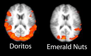
FKF Applied Research and the UCLA Ahmanson Lovelace Brain Mapping Center have released their Second Annual Ranking of the most effective Super Bowl ads using fMRI (functional Magnetic Resonance Imaging) brain imaging. Many of the Super Bowl ads stoked regions of the brain associated with anxiety, including the amygdala.
Compared to last year’s ads there was much more anxiety, and far less positive emotion in these highly touted commercials. “This clearly was the year of the amygdala, the brain’s ‘threat detector,” said Dr. Joshua Freedman UCLA Clinical Assistant Professor of Psychiatry and a co-founder of FKF Applied Research. “Much of the anxiety seemed caused by violence, but was also rooted in economic fears. The Nationwide ad had a spike when Kevin Federline was revealed to be working in fast food, and also when the GM robot turned out to be OK but afraid for its job.”
FKF Applied Research and Dr. Marco Iacoboni’s group at the UCLA Ahmanson Lovelace Brain Mapping Center recruited men and women ages 18-34 to watch this year’s Super Bowl ads. The subjects viewed the ads while in UCLA’s high-field fMRI scanner, which monitors the activity in their brains.
One of the more interesting abstracts from last year’s ISMRM in Seattle has now been published as a full manuscript:
Propeller EPI in the other direction
A new propeller EPI pulse sequence with reduced sensitivity to field inhomogeneities is proposed. Image artifacts such as blurring due to Nyquist ghosting and susceptibility gradients are investigated and compared with those obtained in previous propeller EPI studies. The proposed propeller EPI sequence uses a readout that is played out along the short axis of the propeller blade, orthogonal to the readout used in previous propeller methods. In contrast to long-axis readout propeller EPI, this causes the echo spacing between two consecutive phase-encoding (PE) lines to decrease, which in turn increases the k-space velocity in this direction and hence the pseudo-bandwidth. Long- and short-axis propeller EPI, and standard single-shot EPI sequences were compared on phantoms and a healthy volunteer. Diffusion-weighted imaging (DWI) was also performed on the volunteer. Short-axis propeller EPI produced considerably fewer image artifacts compared to the other two sequences. Further, the oblique blades for the long-axis propeller EPI were also prone to one order of magnitude higher residual ghosting than the proposed short-axis propeller EPI.
Skare S et al, Magn Reson Med. 2006 Jun;55(6):1298. PMID: 16676335
MRI, as an imaging modality, offers tremendous soft-tissue contrast without any use of contrast agents. Even beyond anatomic scans with T1, T2 or proton density weighting, other methods like arterial spin labeling or diffusion imaging exist which leverage both physics and physiology to tease out new information in a scan, making MRI the single most versatile non-invasive imaging method in the medical arsenal.
However, the use of paramagnetic contrast agents (usually administered via IV) is still an integral part of a routine MR exam, for good reason: contrast in MR simply gives us another lever with which we can manipulate the system, providing much more anatomic and functional information. To that end, the Nephrogenic Systemic Fibrosis (NSF) advisory from the FDA is of grave concern. For the vast majority of patients, gadolinium-based contrast agents are perfectly safe, but there still is a need to look for improvement.
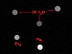
There are some interesting signs in the literature that point to where contrast agents are headed. For one thing, superparamagnetic iron oxide-based (SPIO) contrast agents are already being used, like Feridex®, primarily in abdominal imaging of liver or spleen. These agents’ particles are on the order of 50 nm. However, there’s a new report from the National Institute of Standards and Technology (NIST) about nano-scale iron agents, called “molecular nanomagnets“. Quoting from the NIST press release,
…iron-containing magnets just two nanometers wide, dissolved in water, do provide reasonable contrast in non-clinical MRI images—as long as the nanomagnet concentration is below a certain threshold. (A nanometer is one billionth of a meter.) Previous studies by other research groups had reached conflicting conclusions on the utility of molecular nanomagnets for MRI, but without accounting for concentration. NIST scientists, making novel magnetic measurements, were able to monitor the molecules’ decomposition and magnetic properties as the composition was varied.
The article includes the following image, with caption:
This test image shows what happens when nanomagnets are used to alter the nuclear properties of hydrogen in water, increasing brightness (bright spots below left and center) compared to deionized water (above).
These magnets are a single molecule, less than 5 nm in size. The potential for increased contrast effectiveness, as well as being able to label other biocompounds with them for true molecular imaging, is enormous.
Leveraging nanotech isn’t limited to iron-based agents, however. Even the workhorse Gadolinium-based agents are getting a nanoscale refresh. For example, superparamagnetic gadonanotubes have also been shown to have improved contrast properties. The link is to a paper from August 2005 at Rice University, whose abstract is:
We report the nanoscale loading and confinement of aquated Gd3+n-ion clusters within ultra-short single-walled carbon nanotubes (US-tubes); these Gd3+n@US-tube species are linear superparamagnetic molecular magnets with Magnetic Resonance Imaging (MRI) efficacies 40 to 90 times larger than any Gd3+-based contrast agent (CA) in current clinical use.
There have been a spate of other published works in the literature in this direction as well, of course. It’s clear that there is a lot of ground to be gained yet in terms of optimizing these new agents and bringing them to market. But assuming that they do get there, the nanoscale revolution will certainly leave its mark on MRI as a field. I wouldn’t be surprised to see nanotech contrast agents in the market within 5 years, or even less.