(In addition to MRI and medical physics, it’s worth keeping an open mind and keeping tabs on various other branches of physics and science. To that end, I’ll highlight interesting papers or research that strikes my fancy from time to time.)
Eric Berger aka SciGuy, a science columnist at the Houston Chronicle, points to a new paper in Science that introduces new “metamaterials” which can manipulate light, which are easy to fabricate (in principle). Eric makes the analogy to this being as much a game-changer as lasers were when they were invented almost exactly 50 years ago.
The self-assembly of colloids is an alternative to top-down processing that enables the fabrication of nanostructures. We show that self-assembled clusters of metal-dielectric spheres are the basis for nanophotonic structures. By tailoring the number and position of spheres in close-packed clusters, plasmon modes exhibiting strong magnetic and Fano-like resonances emerge. The use of identical spheres simplifies cluster assembly and facilitates the fabrication of highly symmetric structures. Dielectric spacers are used to tailor the interparticle spacing in these clusters to be approximately 2 nanometers. These types of chemically synthesized nanoparticle clusters can be generalized to other two- and three-dimensional structures and can serve as building blocks for new metamaterials.
and here’s a link to the full text of the article. As with lasers when they were first introduced, it’s a challenge to the imagination to envision how this might be used or applied. What possible medical imaging applications could this be exploited for? That’s the billion dollar question 🙂
MRI, as an imaging modality, offers tremendous soft-tissue contrast without any use of contrast agents. Even beyond anatomic scans with T1, T2 or proton density weighting, other methods like arterial spin labeling or diffusion imaging exist which leverage both physics and physiology to tease out new information in a scan, making MRI the single most versatile non-invasive imaging method in the medical arsenal.
However, the use of paramagnetic contrast agents (usually administered via IV) is still an integral part of a routine MR exam, for good reason: contrast in MR simply gives us another lever with which we can manipulate the system, providing much more anatomic and functional information. To that end, the Nephrogenic Systemic Fibrosis (NSF) advisory from the FDA is of grave concern. For the vast majority of patients, gadolinium-based contrast agents are perfectly safe, but there still is a need to look for improvement.
There are some interesting signs in the literature that point to where contrast agents are headed. For one thing, superparamagnetic iron oxide-based (SPIO) contrast agents are already being used, like Feridex®, primarily in abdominal imaging of liver or spleen. These agents’ particles are on the order of 50 nm. However, there’s a new report from the National Institute of Standards and Technology (NIST) about nano-scale iron agents, called “molecular nanomagnets“. Quoting from the NIST press release,
…iron-containing magnets just two nanometers wide, dissolved in water, do provide reasonable contrast in non-clinical MRI images—as long as the nanomagnet concentration is below a certain threshold. (A nanometer is one billionth of a meter.) Previous studies by other research groups had reached conflicting conclusions on the utility of molecular nanomagnets for MRI, but without accounting for concentration. NIST scientists, making novel magnetic measurements, were able to monitor the molecules’ decomposition and magnetic properties as the composition was varied.
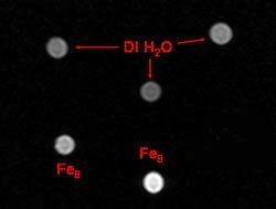
The article includes the following image, with caption:
This test image shows what happens when nanomagnets are used to alter the nuclear properties of hydrogen in water, increasing brightness (bright spots below left and center) compared to deionized water (above).
These magnets are a single molecule, less than 5 nm in size. The potential for increased contrast effectiveness, as well as being able to label other biocompounds with them for true molecular imaging, is enormous.
Leveraging nanotech isn’t limited to iron-based agents, however. Even the workhorse Gadolinium-based agents are getting a nanoscale refresh. For example, superparamagnetic gadonanotubes have also been shown to have improved contrast properties. The link is to a paper from August 2005 at Rice University, whose abstract is:
We report the nanoscale loading and confinement of aquated Gd3+n-ion clusters within ultra-short single-walled carbon nanotubes (US-tubes); these Gd3+n@US-tube species are linear superparamagnetic molecular magnets with Magnetic Resonance Imaging (MRI) efficacies 40 to 90 times larger than any Gd3+-based contrast agent (CA) in current clinical use.
There have been a spate of other published works in the literature in this direction as well, of course. It’s clear that there is a lot of ground to be gained yet in terms of optimizing these new agents and bringing them to market. But assuming that they do get there, the nanoscale revolution will certainly leave its mark on MRI as a field. I wouldn’t be surprised to see nanotech contrast agents in the market within 5 years, or even less.