An MRI unit exploded last year at Peninsula Regional Medical Center in Maryland. This was actually caught on film by a local TV crew!
Neat.
Just for fun, here’s a bonus video of a chair stuck in a magnet. That looks like a lot of work.
a celebration of science fiction, anime, and geek culture

An MRI unit exploded last year at Peninsula Regional Medical Center in Maryland. This was actually caught on film by a local TV crew!
Neat.
Just for fun, here’s a bonus video of a chair stuck in a magnet. That looks like a lot of work.
news flash: emotions sometime trump rational thought! Shocking, I know. Though I was intrigued at how the fMRI paradigm in this case provides a neat empirical example for why prisoner’s dilemma models don’t translate well into real-world practice:
A classic economic example is the “ultimatum game,” in which one participant gets 10 $1 bills (or loonies, in Canada). He chooses how many to offer to a second participant. If she accepts the offer, the money is split the way the first participant suggested; if she rejects the offer, nobody gets anything.
Logically, the first participant can maximize his money by offering a single dollar, because logically the second participant should accept that as being better than nothing. In real life, however, the second participant, if offered only a dollar or two, almost always rejects the offer.
Functional MRI scans of brain activity show that a low offer stimulates an area associated with negative emotions, including anger and disgust. It seems the second participant would rather punish the first participant for making such an insulting offer than make an easy buck. And usually, the person making the offer understands this and offers something close to an even split, averaging about $4.
I don’t really see why the above reasonable decision-making process is inherently non-rational or “emotional” though. Doesn’t it make good rational sense to “punish” someone making a lowball offer, so they are motivated to offer you more up front?
An editorial in the Journal of Nuclear Medicine (J Nucl Med. 2007 Mar;48(3):331. PMID: 17332606) argues that combination PET/MRI systems are the future and will supplant PET/CT:
In a number of ways, the path to PET/MRI has been reverse of that to PET/CT. The first PET/CT design emerged from industry–academia collaboration and was a prototype for human clinical use that eventually stimulated a commercial response and led to the development of PET/CT for imaging small animals. In contrast, PET/MRI began with the small-animal design and then, over a decade later, the first PET/MRI brain images were acquired on a dedicated prototype system, following an impressive industrial backing that far exceeded that of the early PET/CT developments.
[…]
A mere 2 y after the advent of commercial PET/CT, Johannes Czernin from UCLA, at the 2003 annual DGN meeting, commented that “PET/CT is a technical evolution that has led to a medical revolution.” Today, at the dawn of PET/MRI, it may be said that “PET/MRI is a medical evolution based on a technical revolution.” Although PET/CT appears to have replaced stand-alone PET for most oncologic indications, it is reasonable to assume that PET/MRI will be the preferred imaging option for neurologic and central nervous system indications. Without doubt, such dual-modality combinations are here to stay because they incorporate the diagnostic power of PET. Thus, PET/CT and PET/MRI, by virtue of their combined anatometabolic imaging, will lead to a “new-clear” medicine and the demise of “unclear” medicine.
That’s “not safe for work”, not “national science foundation” up there in the acronym. It was surely inevitable that this amazing, subtle and elegant technology would eventually be applied to more scatological pursuits. The following paper is a classic in this genre.
Magnetic resonance imaging of male and female genitals during coitus and female sexual arousal.
OBJECTIVE: To find out whether taking images of the male and female genitals during coitus is feasible and to find out whether former and current ideas about the anatomy during sexual intercourse and during female sexual arousal are based on assumptions or on facts. DESIGN: Observational study. SETTING: University hospital in the Netherlands. METHODS: Magnetic resonance imaging was used to study the female sexual response and the male and female genitals during coitus. Thirteen experiments were performed with eight couples and three single women. RESULTS: The images obtained showed that during intercourse in the “missionary position” the penis has the shape of a boomerang and 1/3 of its length consists of the root of the penis. During female sexual arousal without intercourse the uterus was raised and the anterior vaginal wall lengthened. The size of the uterus did not increase during sexual arousal. CONCLUSION: Taking magnetic resonance images of the male and female genitals during coitus is feasible and contributes to understanding of anatomy.
Schultz et al, BMJ. 1999 Dec 18-25;319(7225):1596-600. PMID 10600954.
I am sure that this experiment was initially conceived by grad students:
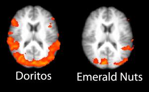
FKF Applied Research and the UCLA Ahmanson Lovelace Brain Mapping Center have released their Second Annual Ranking of the most effective Super Bowl ads using fMRI (functional Magnetic Resonance Imaging) brain imaging. Many of the Super Bowl ads stoked regions of the brain associated with anxiety, including the amygdala.
Compared to last year’s ads there was much more anxiety, and far less positive emotion in these highly touted commercials. “This clearly was the year of the amygdala, the brain’s ‘threat detector,” said Dr. Joshua Freedman UCLA Clinical Assistant Professor of Psychiatry and a co-founder of FKF Applied Research. “Much of the anxiety seemed caused by violence, but was also rooted in economic fears. The Nationwide ad had a spike when Kevin Federline was revealed to be working in fast food, and also when the GM robot turned out to be OK but afraid for its job.”
FKF Applied Research and Dr. Marco Iacoboni’s group at the UCLA Ahmanson Lovelace Brain Mapping Center recruited men and women ages 18-34 to watch this year’s Super Bowl ads. The subjects viewed the ads while in UCLA’s high-field fMRI scanner, which monitors the activity in their brains.
One of the more interesting abstracts from last year’s ISMRM in Seattle has now been published as a full manuscript:
Propeller EPI in the other direction
A new propeller EPI pulse sequence with reduced sensitivity to field inhomogeneities is proposed. Image artifacts such as blurring due to Nyquist ghosting and susceptibility gradients are investigated and compared with those obtained in previous propeller EPI studies. The proposed propeller EPI sequence uses a readout that is played out along the short axis of the propeller blade, orthogonal to the readout used in previous propeller methods. In contrast to long-axis readout propeller EPI, this causes the echo spacing between two consecutive phase-encoding (PE) lines to decrease, which in turn increases the k-space velocity in this direction and hence the pseudo-bandwidth. Long- and short-axis propeller EPI, and standard single-shot EPI sequences were compared on phantoms and a healthy volunteer. Diffusion-weighted imaging (DWI) was also performed on the volunteer. Short-axis propeller EPI produced considerably fewer image artifacts compared to the other two sequences. Further, the oblique blades for the long-axis propeller EPI were also prone to one order of magnitude higher residual ghosting than the proposed short-axis propeller EPI.
Skare S et al, Magn Reson Med. 2006 Jun;55(6):1298. PMID: 16676335
MRI, as an imaging modality, offers tremendous soft-tissue contrast without any use of contrast agents. Even beyond anatomic scans with T1, T2 or proton density weighting, other methods like arterial spin labeling or diffusion imaging exist which leverage both physics and physiology to tease out new information in a scan, making MRI the single most versatile non-invasive imaging method in the medical arsenal.
However, the use of paramagnetic contrast agents (usually administered via IV) is still an integral part of a routine MR exam, for good reason: contrast in MR simply gives us another lever with which we can manipulate the system, providing much more anatomic and functional information. To that end, the Nephrogenic Systemic Fibrosis (NSF) advisory from the FDA is of grave concern. For the vast majority of patients, gadolinium-based contrast agents are perfectly safe, but there still is a need to look for improvement.
There are some interesting signs in the literature that point to where contrast agents are headed. For one thing, superparamagnetic iron oxide-based (SPIO) contrast agents are already being used, like Feridex®, primarily in abdominal imaging of liver or spleen. These agents’ particles are on the order of 50 nm. However, there’s a new report from the National Institute of Standards and Technology (NIST) about nano-scale iron agents, called “molecular nanomagnets“. Quoting from the NIST press release,
…iron-containing magnets just two nanometers wide, dissolved in water, do provide reasonable contrast in non-clinical MRI images—as long as the nanomagnet concentration is below a certain threshold. (A nanometer is one billionth of a meter.) Previous studies by other research groups had reached conflicting conclusions on the utility of molecular nanomagnets for MRI, but without accounting for concentration. NIST scientists, making novel magnetic measurements, were able to monitor the molecules’ decomposition and magnetic properties as the composition was varied.
The article includes the following image, with caption:
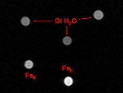
These magnets are a single molecule, less than 5 nm in size. The potential for increased contrast effectiveness, as well as being able to label other biocompounds with them for true molecular imaging, is enormous.
Leveraging nanotech isn’t limited to iron-based agents, however. Even the workhorse Gadolinium-based agents are getting a nanoscale refresh. For example, superparamagnetic gadonanotubes have also been shown to have improved contrast properties. The link is to a paper from August 2005 at Rice University, whose abstract is:
We report the nanoscale loading and confinement of aquated Gd3+n-ion clusters within ultra-short single-walled carbon nanotubes (US-tubes); these Gd3+n@US-tube species are linear superparamagnetic molecular magnets with Magnetic Resonance Imaging (MRI) efficacies 40 to 90 times larger than any Gd3+-based contrast agent (CA) in current clinical use.
There have been a spate of other published works in the literature in this direction as well, of course. It’s clear that there is a lot of ground to be gained yet in terms of optimizing these new agents and bringing them to market. But assuming that they do get there, the nanoscale revolution will certainly leave its mark on MRI as a field. I wouldn’t be surprised to see nanotech contrast agents in the market within 5 years, or even less.
Ars Technica has a nice summary of a recent paper in PLoS that attempted to assess the quality of the peer review process. From the Ars summary:
To examine what makes a good reviewer, they took advantage of the journal Annals of Emergency Medicine, which has maintained a detailed database of reviewers and post-review ratings (on a five-point scale) of their work, performed by the editors of the journal. The researchers contacted the reviewers and surveyed them about various factors that might contribute to skill in the process. A diverse set of 306 reviewers who had performed a total of nearly 3,000 reviews were used as the data set.
[…]
In news that may be disturbing for journal editors everywhere, very few factors leapt out as having a consistent and significant correlation with the quality of a review, although some factors did have strong correlations in individual tests. The only positive factors linked to quality of reviews were age (younger reviewers were better) and working at an academic hospital. Ironically, service on an Institutional Review Board, which evaluates and approves experiments on humans, consistently correlated with lower-quality peer reviews. Even these factors, however, were only slightly better than random at predicting review quality.
Ultimately the peer review process is always going to have a subjective component to it, since the processes of intuition and patterning that are fundamental to scientific insight and understanding are not really very deterministic. But there’s another possible reason why the study failed to find strong correlates of review quality; the very assessment of quality itself is equally subjective. I personally believe that the peer review system is like democracy – far from ideal but better than anything else out there. The best way to ensure general quality is to ensure that a maximum number of scientists in a given field participate in the process. Perhaps one way to achieve this would be to extend reviewer privileges to graduate students who have passed oral qualifiers?
Journal article citation: Callaham ML, Tercier J (2007) The Relationship of Previous Training and Experience of Journal Peer Reviewers to Subsequent Review Quality. PLoS Med 4(1): e40 doi:10.1371/journal.pmed.0040040
Related article: Kotchen TA, Lindquist T, Miller Sostek A, Hoffmann R, Malik K, Stanfield B. Outcomes of National Institutes of Health peer review of clinical grant applications. J Investig Med. 2006 Jan;54(1):13-9. PMID: 16409886
The FDA has an updated Public Health Advisory on the safety of gadolinium-based MRI contrast agents. In a nutshell, patients with any sort of renal disease or otherwise compromised kidney function are at high risk of developing Nephrogenic Systemic Fibrosis (NSF) or Nephrogenic Fibrosing Dermopathy (NFD) if they receive gadolinium contrast agents for MRI imaging. There seems to be no risk for patients without compromised kidney function.
Continue reading “FDA Advisory on Gadolinium contrast agents”